KERATOCONUS

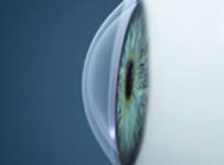
Keratoconus is a disorder of the cornea that leads to progressive changes in its shape and structure. Over time, the cornea becomes increasingly thinner and steeper. These changes result in the formation of high levels of irregular astigmatism and myopia.

Normal cornea
Cross-sectional view of a normal cornea, note the round nature of the cornea, which is symmetric in each direction.

Keratoconus
Cross-sectional view of a cornea displaying classic keratoconus changes. The cornea is bowed in a cone shape and the tissue has become thinned out.
As the cornea becomes increasingly thinner, it can lead to corneal edema (swelling) and bullous keratopathy (corneal blisters). Ultimately, when the swelling subsides, the cornea is left permanently scarred.
Incidence & Cause
In the general population it occurs in approximately 1 in 2,500 people. At this time the exact cause of Keratoconus is not fully understood, but it has been suggestive to be multifactorial in nature. It has been associated with a genetic predisposition, chronic allergic conjunctivitis, eye rubbing, and an abnormal formation of collagen, the major structural component of the cornea.
Treatment
The mainstay treatment for Keratoconus has been aimed at correction of high levels of refractive errors (astigmatism and myopia) that cause poor vision. In the earlier stages of the disease, one’s vision can be corrected with glasses or specialty contact lenses. Unfortunately, as the cone progresses these options become incapable of correcting one’s vision. In the past surgical treatment for this disease had centered upon intrastromal corneal ring segments (INTACS) and corneal transplantation as the only option for improvement of vision.
Fortunately, now there is a treatment aimed at halting the progression of the disease in the earliest stages. This treatment is known as collagen cross-linking. As of April 2016, the FDA approved the Avedro, Collagen Cross-Linking device. This FDA approved treatment is safe, easy, and offered here at Vision NYC.

COLLAGEN CROSS-LINKING
As of April 2016, the FDA approved the first Collagen Cross-Linking system in the United States. But what does this mean for our patients with keratonocus?
Patients with keratonocus will for the first time have access to an FDA approved procedure to prevent the progression of keratonocus. This procedure has been shown to be safe and effective, and has been widely practiced for many years throughout most of the modern world.
What are some of the main goals of the procedure?
-
To stop the progression of the disease.
-
To allow continued use of glasses or contact lenses to achieve functional vision.
Will I see better after the procedure?
In certain cases, patients may see a mild improvement in their vision. This outcome is not typical, and is not the primary purpose of the procedure. The purpose is to halt the progression of the disease and to prevent the need for corneal transplantation in the future.
How does the Collagen Cross-Linking work?
The cornea is composed of sheets of collagen. In patients with keratoconus the collagen lacks sufficient tensile strength, therefore it is prone to changing shape over time. The normal cornea is round like a sphere. The result of weak collagen is the continued progression of keratoconus and the formation of a steeper cone. This creates high levels of astigmatism, scarring, and blurry vision.

CORNEA TRANSPLANT
Keratoconus is a disorder of the cornea that leads to progressive changes in its shape and structure. Over time, the cornea becomes increasingly thinner and steeper. In more advanced cases, patients with keratoconus can develop central scarring of the cornea. All of these changes result in the formation of high levels of irregular astigmatism and myopia. These refractive errors become increasingly difficult to treat through non-surgical options alone, such as glasses of specialty contact lenses. In advanced cases of keratoconus corneal transplantation is routinely performed to restore one’s sight.

Over the years, more successful visual outcomes have been achieved through advancements in surgical technique as well as increased acceptance of donor tissue. At Vision NYC we perform a thorough examination of your eye and use your previous corneal history to aid us in deciding if transplantation surgery is an option which may offer you improved visual acuity. Restoring one’s sight from successful corneal transplantation is truly a life changing experience and we at Vision NYC hope to make your experience a positive one.
